Surgical Procedures
Surgery, shown one stage at a time
Selected procedures photographed at each stage and titled by the surgeon. Every case is published with the patient's consent.
Laparoscopy + Robotic Surgery · Oct 2026
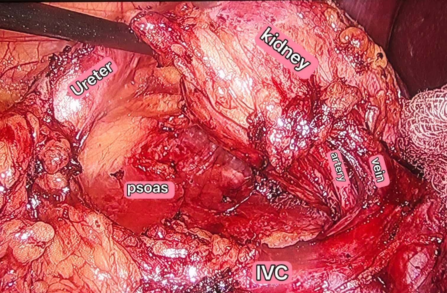
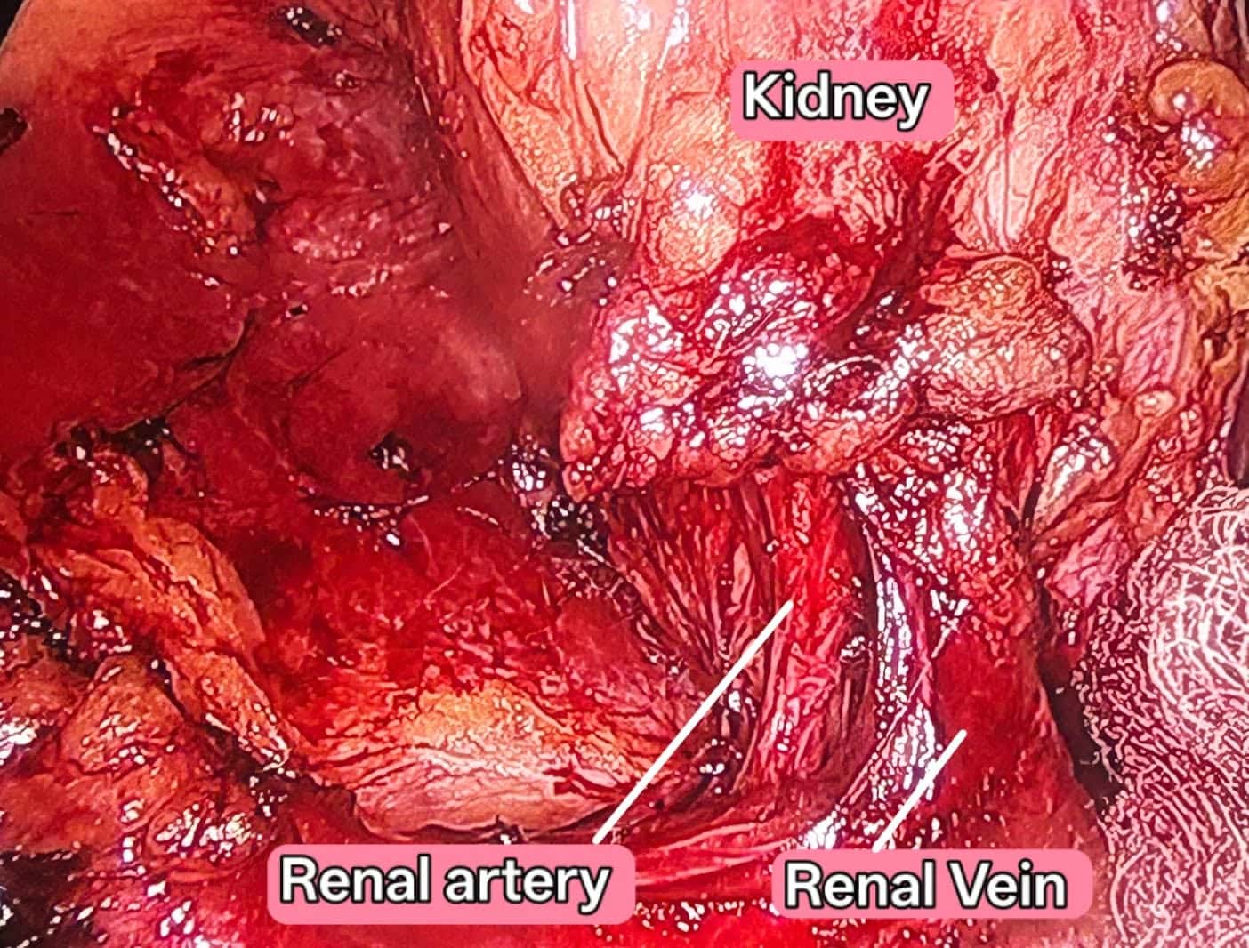
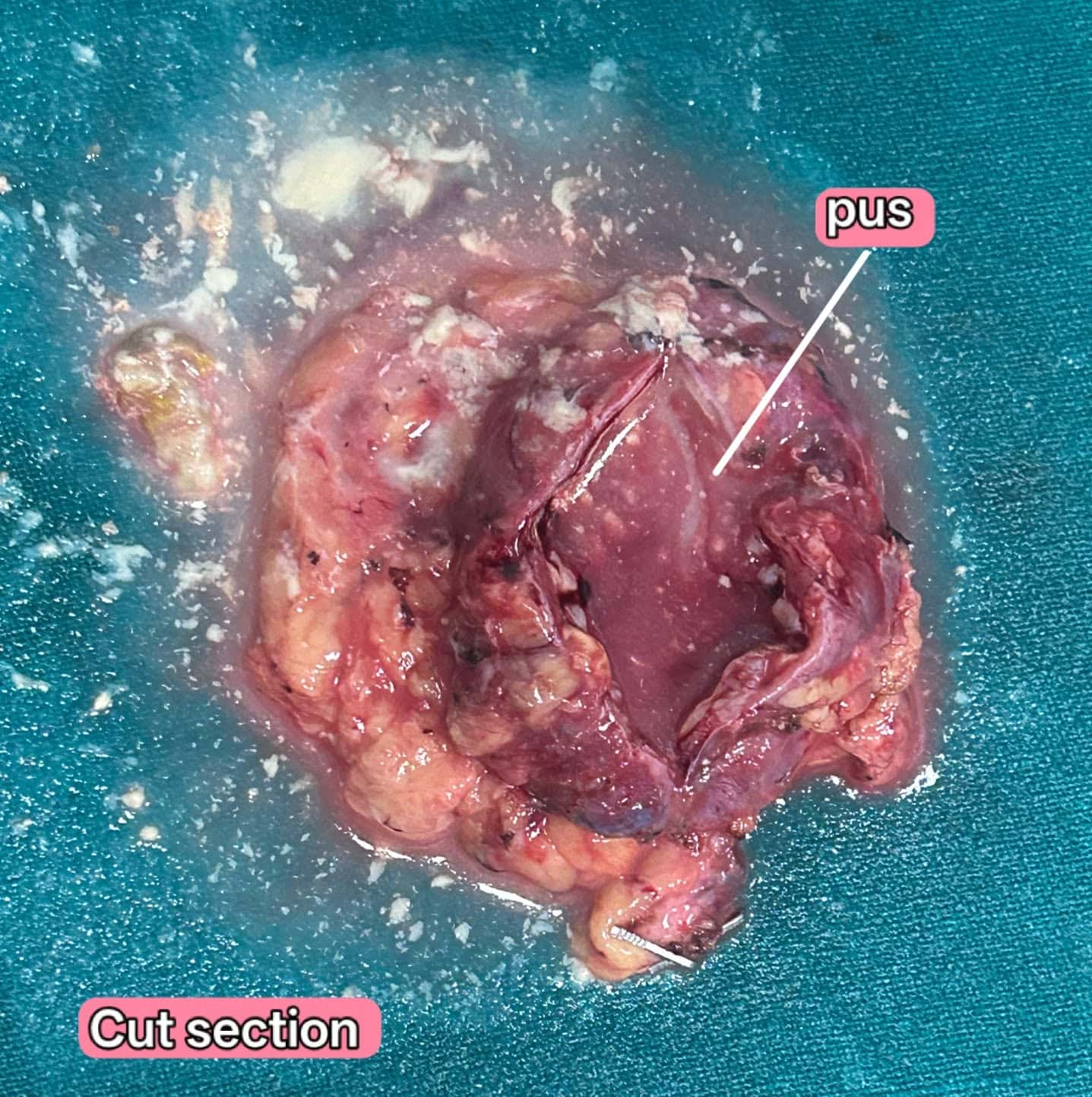
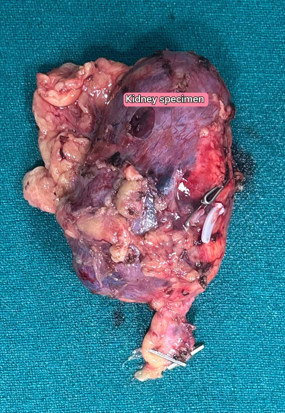
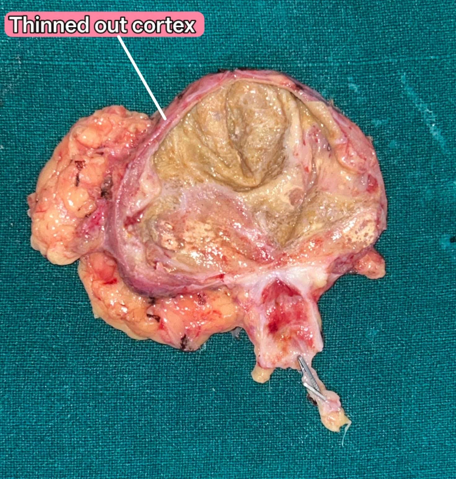
Laparoscopic simple nephrectomy
Laparoscopic simple nephrectomy is the standard, minimally invasive treatment for a non-functioning kidney caused by an upper ureteric calculus (stone) and chronic obstruction or recurrent infection. This patient had a non-functioning kidney due to an upper ureteric calculus with pyonephrosis, leading to recurrent infections.
5 stagesExplore procedure
Laparoscopy + Robotic Surgery · Oct 2026
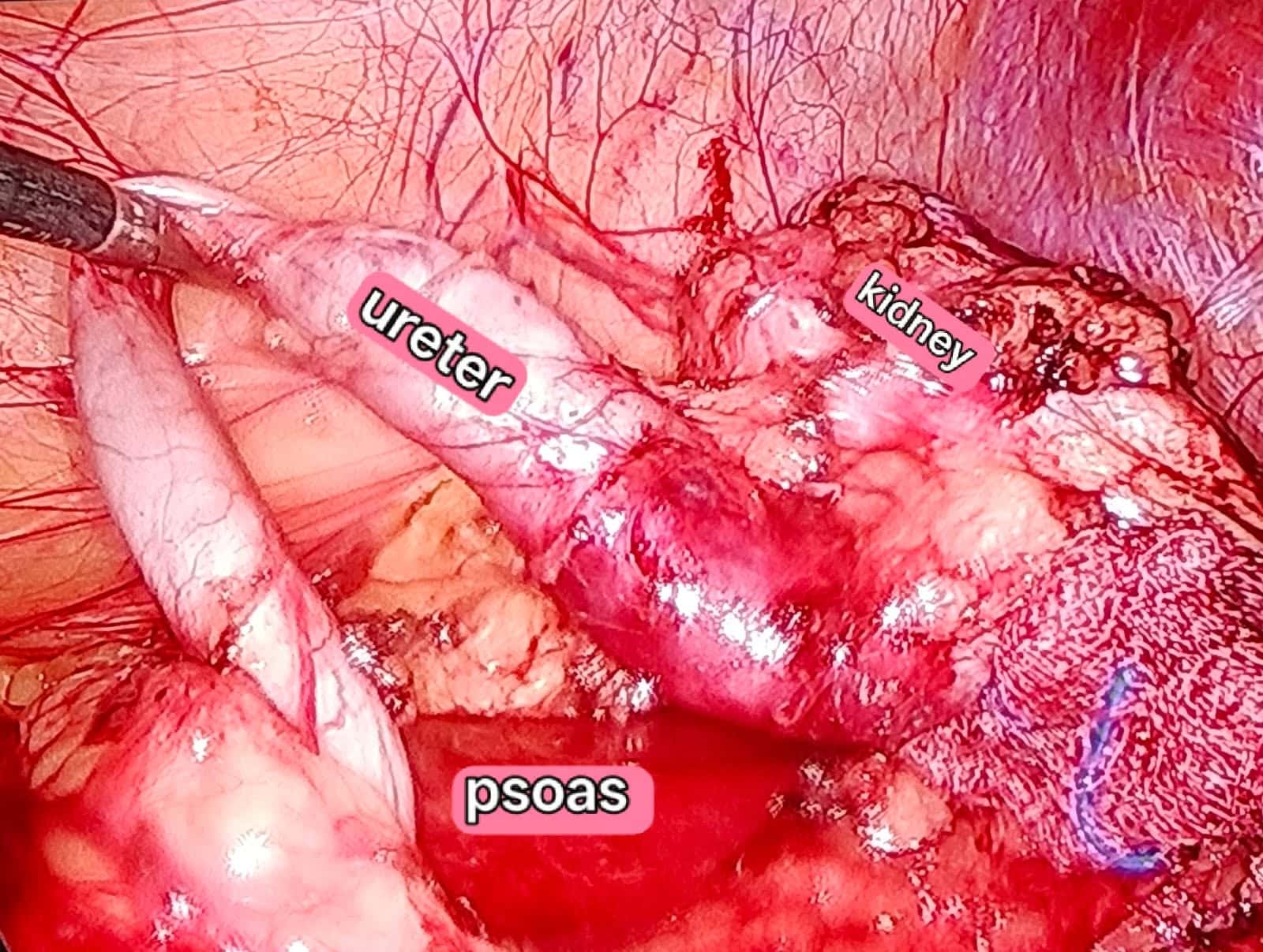
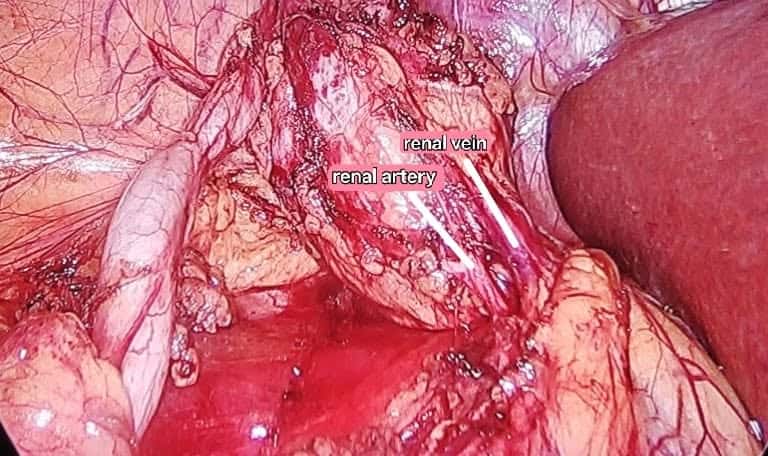
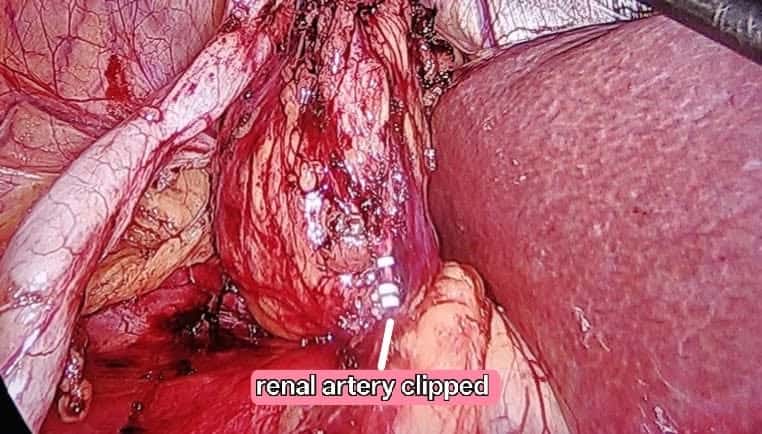
Laparoscopic nephrectomy + Cholecystectomy
This combined approach is typically recommended when a patient presents with coexisting diseases in both the kidney and gallbladder. This combined approach is a safe, highly feasible, and effective strategy that eliminates the need for two separate hospital stays and separate surgeries. This patient had a non-functioning kidney due to a lower ureteric stricture and symptomatic gallstone disease.
11 stagesExplore procedure
Reconstructive Urology · Oct 2026
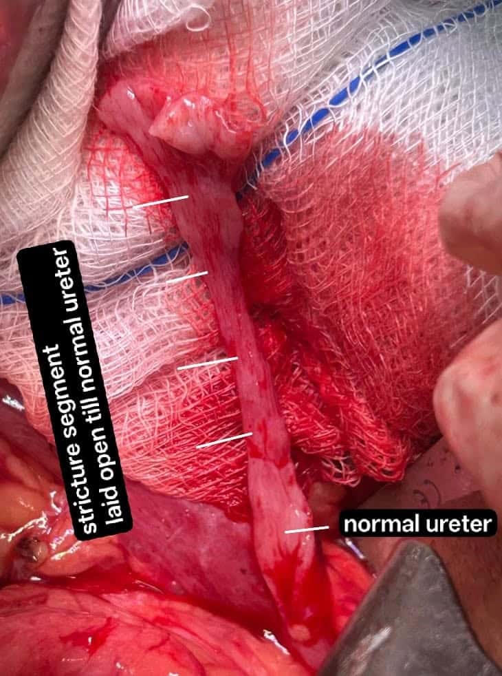
Long segment PUJO
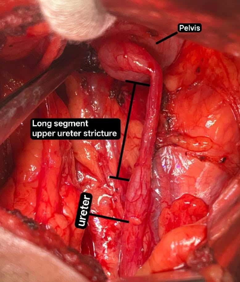
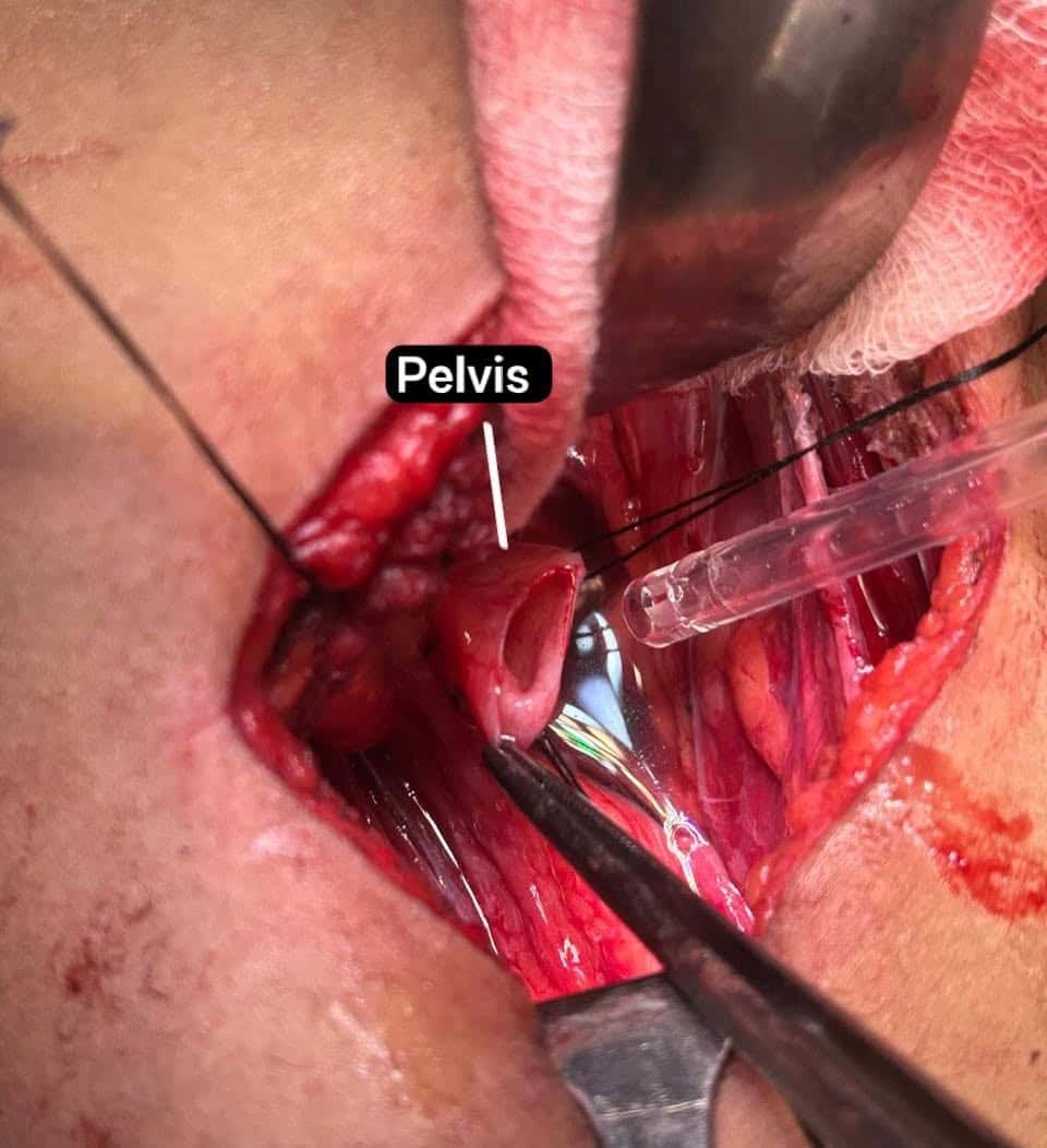
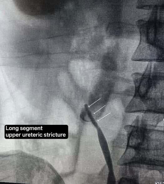
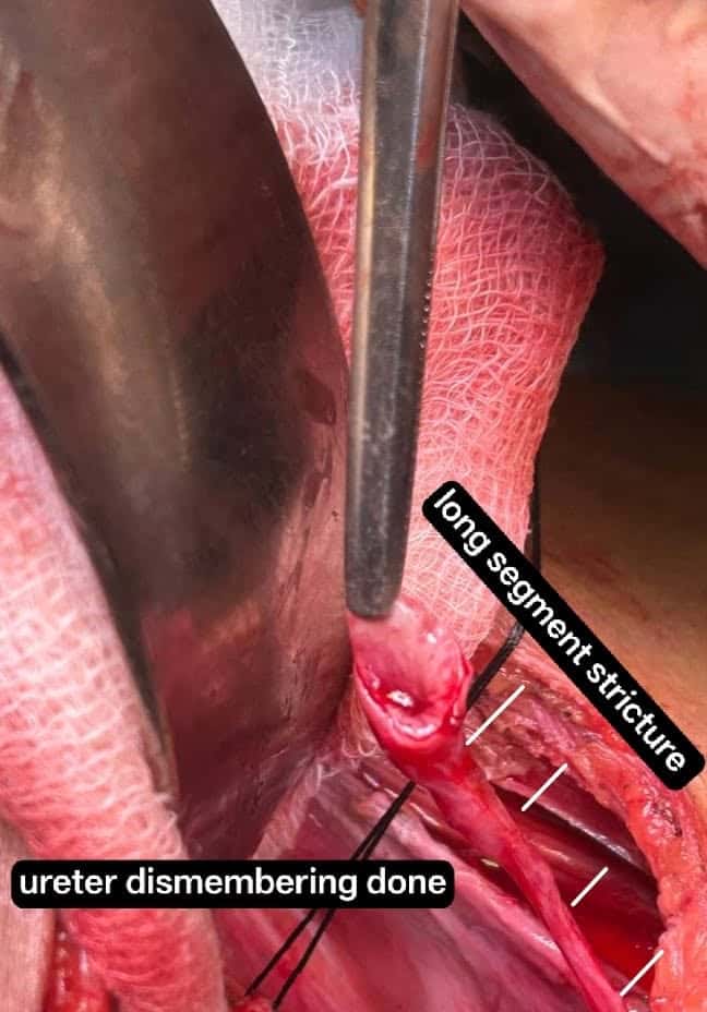
Unlike standard PUJ obstructions, which typically involve a very short, localized segment, a "long segment" stricture spans down the upper ureter. This makes standard surgical fixes more challenging. In such cases, one may not be able to perform a dismembered pyeloplasty; hence, there should always be a backup before you proceed with transection. This can be in the form of a spiral flap (Culp-DeWeerd), a vertical flap (Scardino-Prince), Buccal Mucosa Graft (BMG) ureteroplasty, or ureterocalicostomy. This patient was taken for laparoscopic pyeloplasty. A preoperative retrograde pyelogram was performed because the CT findings were equivocal and showed a long-segment PUJ stricture. The case was converted to open, and luckily, we were able to do A-H dismembered pyeloplasty after good renal mobilisation. In most cases, we do not do RGP's but If non-invasive imaging does not clearly map the stricture, RGP provides real-time visualization of the pelvic-ureteric geometry.
5 stagesExplore procedure
Andrology · Oct 2026
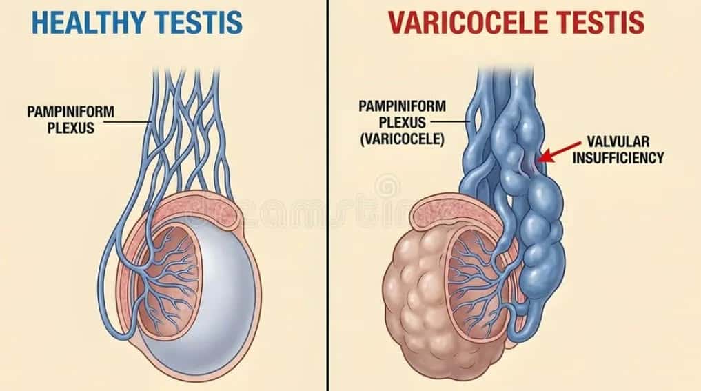
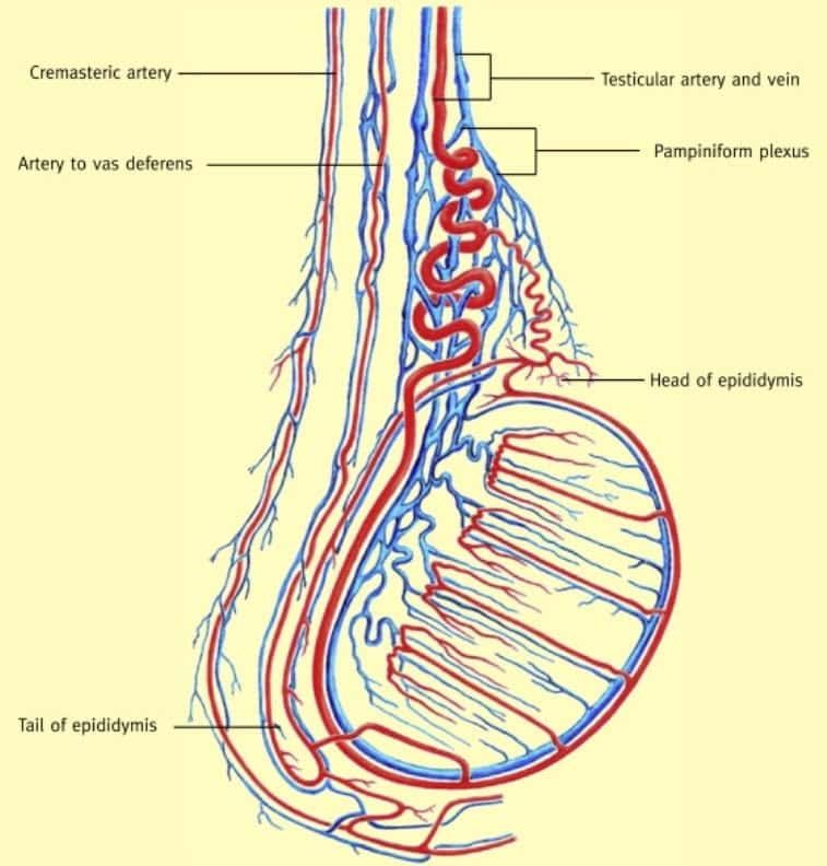
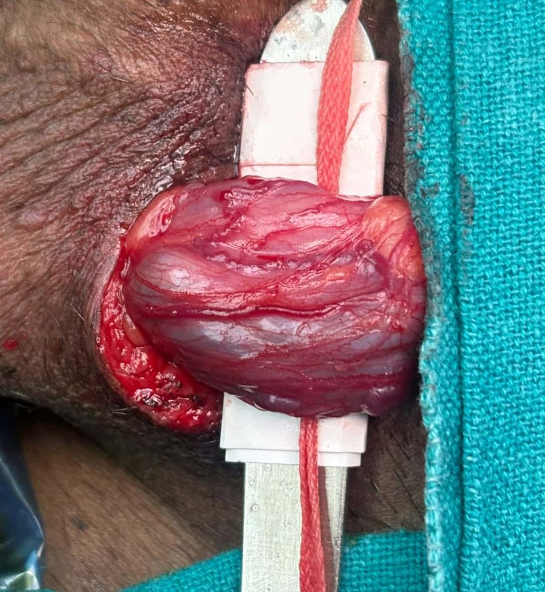
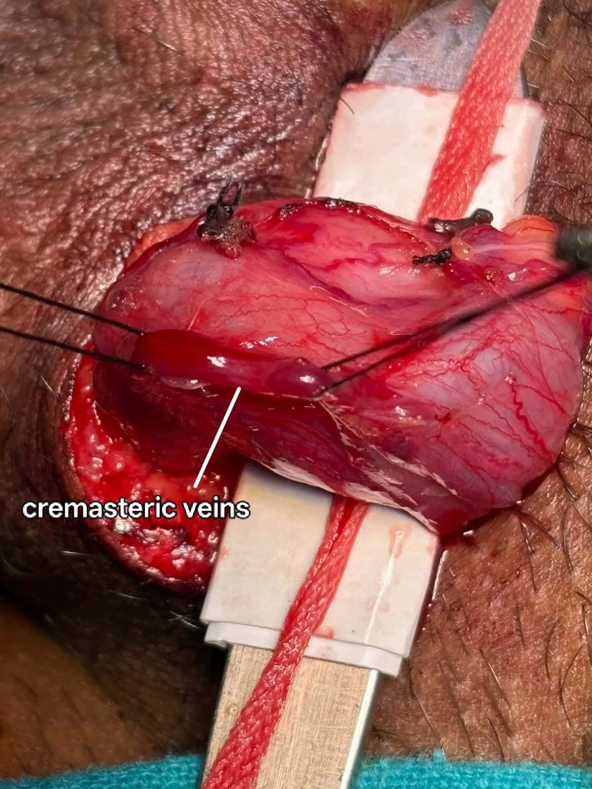
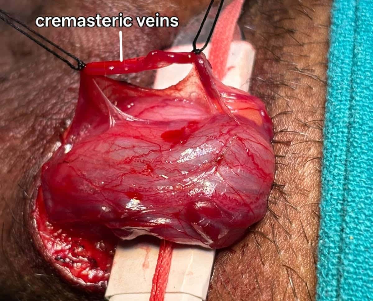
Microscopic variceal ligation
The microsurgical variceal ligation is considered the “gold standard” because of the lowest risk of complications (varicocele recurrence, hydrocele formation [fluid collection around the testicle] and testicular atrophy) and a higher success rate (disappearance of varicocele) The subinguinal approach is more challenging owing to the greater number of vessels (arteries and veins) encountered at this level, compared with the inguinal approach. This patient had a solitary testis with clinical grade III varicocele. Microscopic variceal ligation was done using a microscope of power 10X magnification
9 stagesExplore procedure
Laparoscopy + Robotic Surgery · Oct 2026
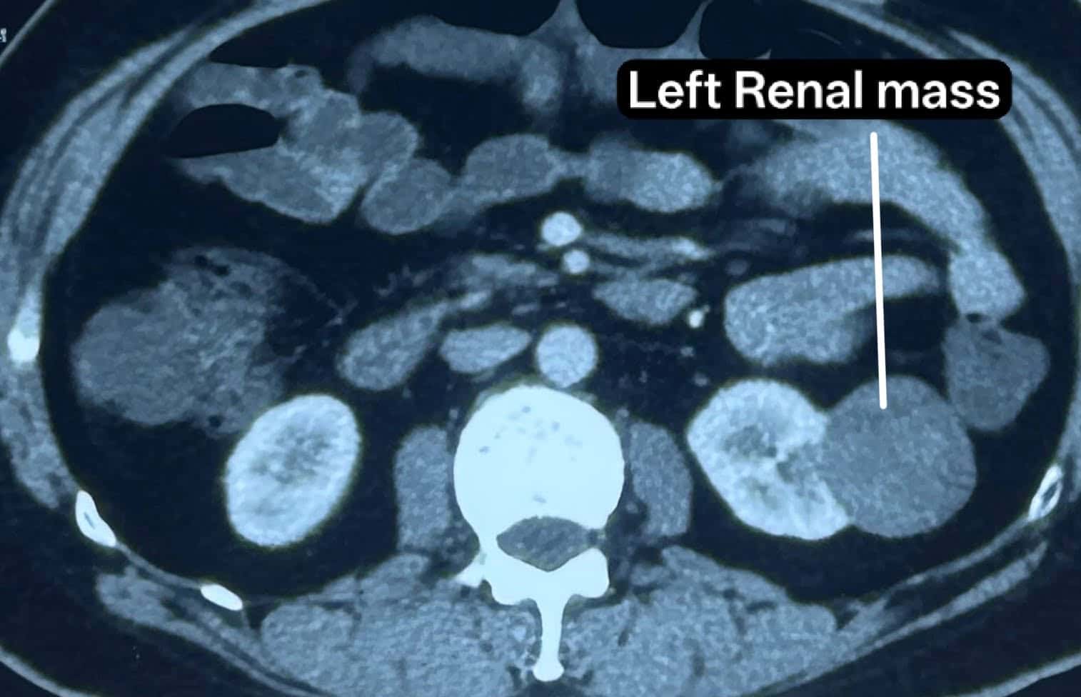
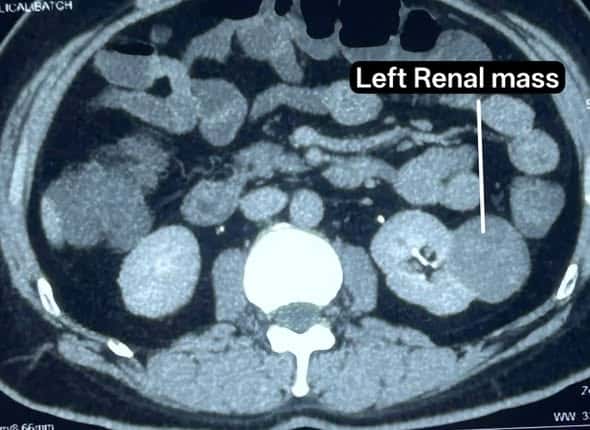
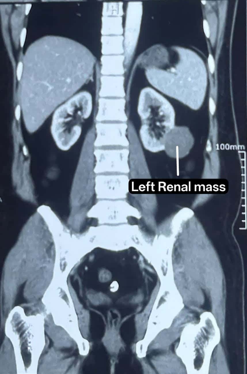
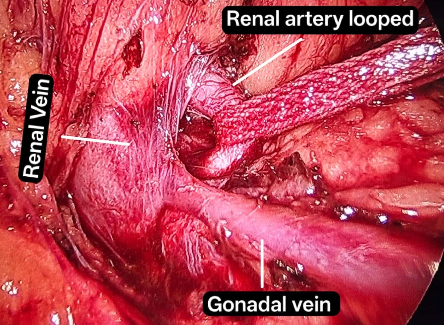
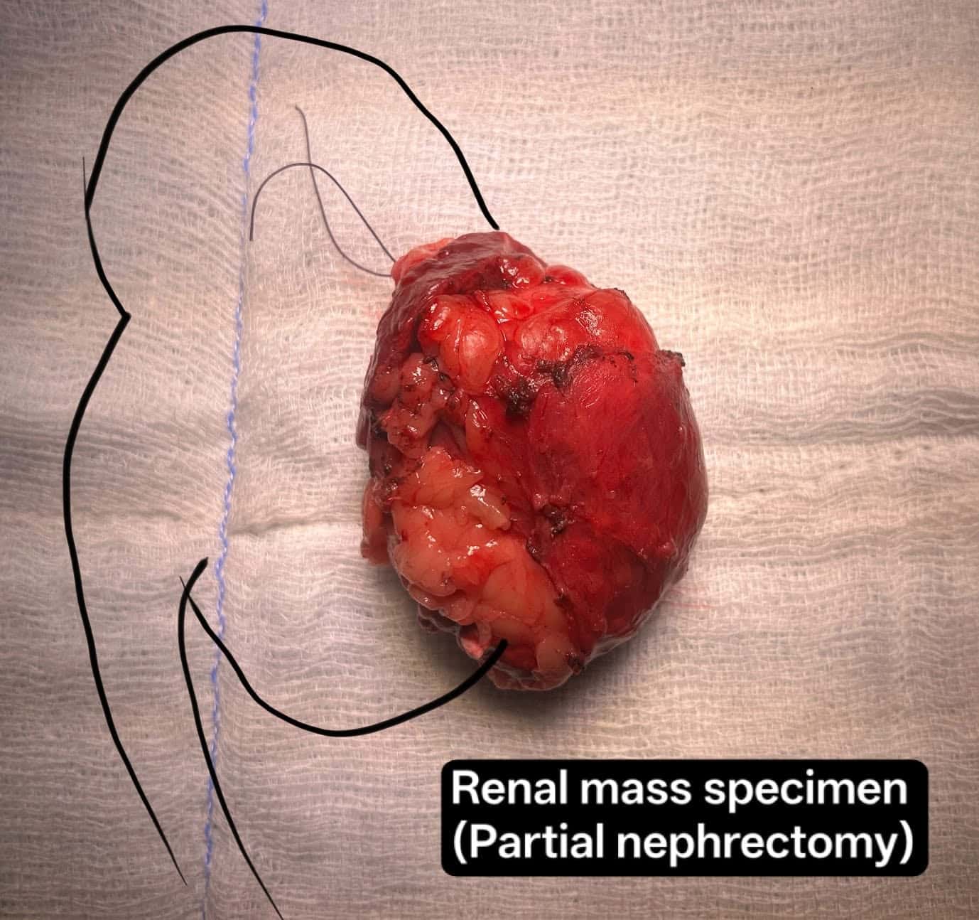
Laparoscopic partial nephrectomy
Laparoscopic partial nephrectomy is one of the first-line surgical techniques for the management of small renal tumors where robotic surgery cannot be done because of financial constraints and unavailability. Laparoscopic partial nephrectomy is very difficult to perform compared to robotic surgery and requires a high degree of expertise. The main advantages are excellent oncologic control together with the preservation of nephron units. Moreover, it implies a shorter length of hospital stay, less postoperative pain, and shorter recovery times for patients. In this case, laparoscopic partial nephrectomy was performed as the patient was not able to afford robotic partial nephrectomy
7 stagesExplore procedure
Laparoscopy + Robotic Surgery · Oct 2026
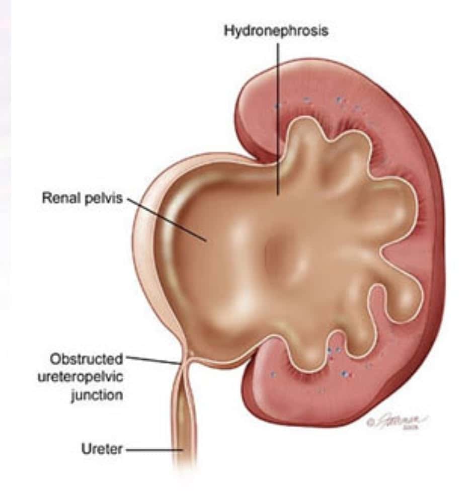
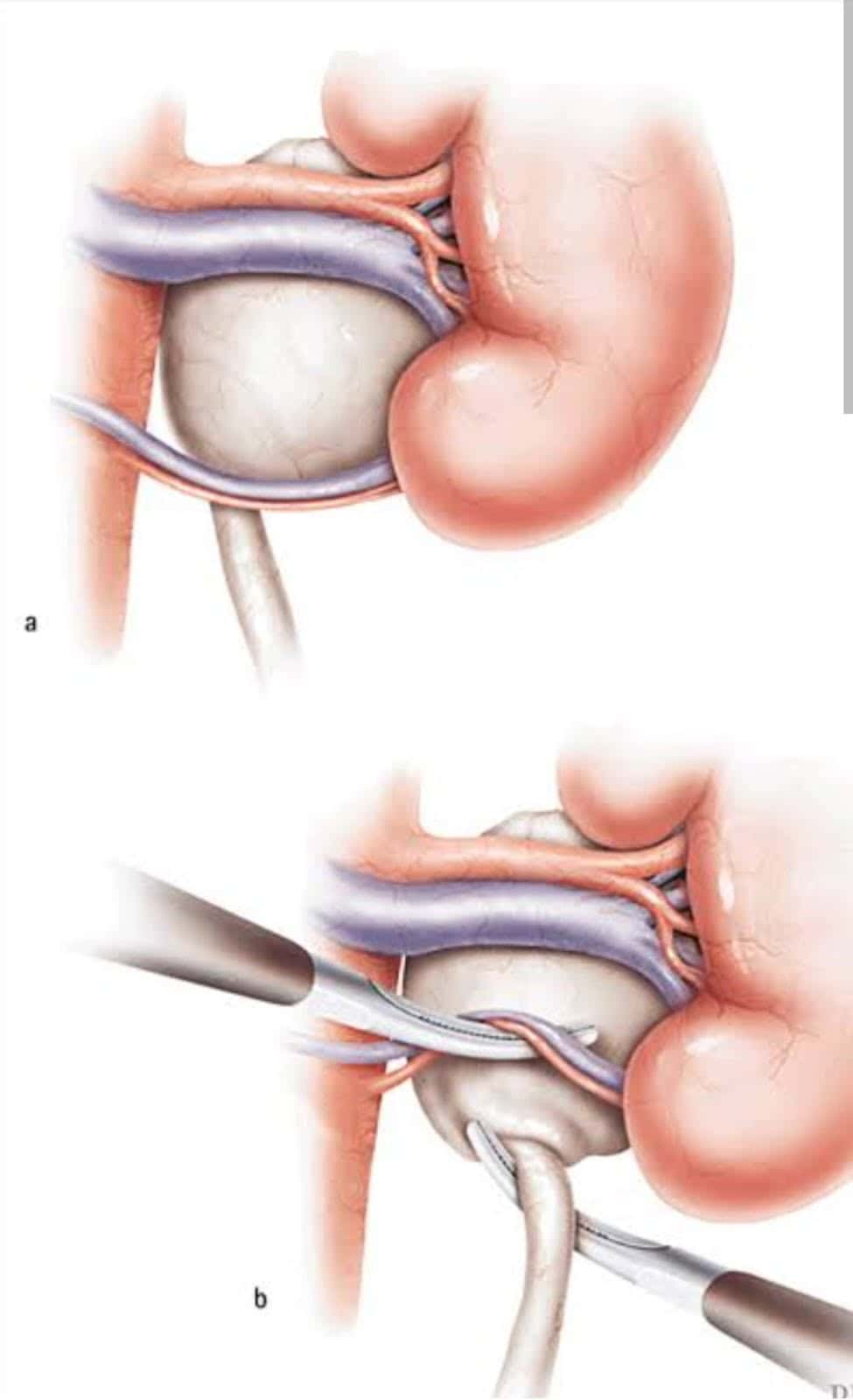
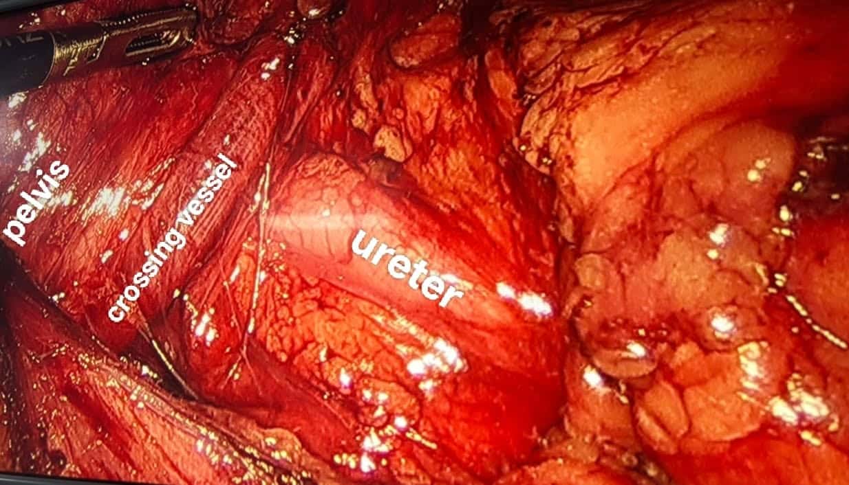
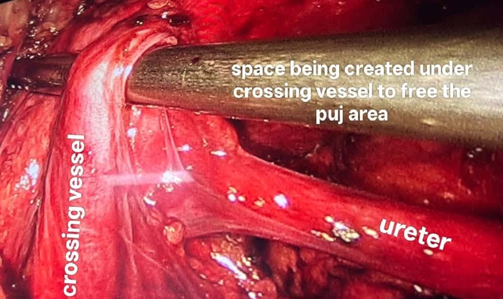
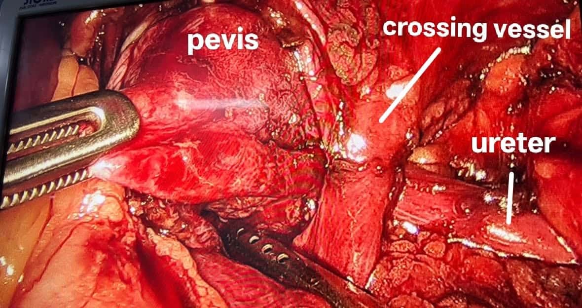
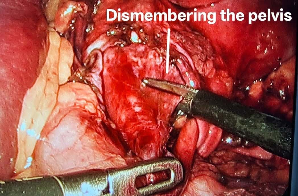
Laparoscopic Pyeloplasty in a case of a crossing vesse
Laparoscopic pyeloplasty with a crossing vessel is technically more demanding than a straightforward UPJ obstruction. The main challenge is safely dissecting the vessel and choosing the correct reconstruction. During dissection, avoid excessive traction, thermal injury, or inadvertent vessel injury, which can cause significant bleeding or compromise renal perfusion. The new uretero-pelvic junction must be tension-free, dependent, and well vascularized. The reconstructed ureter should generally lie anterior to the crossing vessel when a dismembered transposition repair is indicated In this case, a crossing vessel was present, which was supplying the lower pole (only functional parenchyma), was identified intraoperatively and safely preserved
9 stagesExplore procedure





Uro-Oncology
j
kk
14 stagesExplore procedure